RATOC
       RATOC >BONE>bone morphometry
            Analysis service    Bone    Tissue cell reconstruction    Fiber    Otolith    Display options       Particle measurement    Through-hole measurement    4Dkinetic Anlysis      
            Cardiac function measurement        Light scattering tomograph        User support


3D Bone BMD measurement
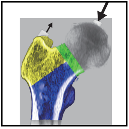
          

Femur measurement
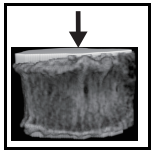
          

Waist, chest, cervical spine
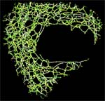
          

bone morphometry
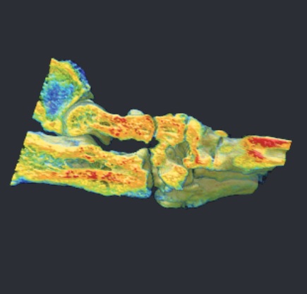
          

Bone mineral density measurement
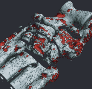
          

Rheumatism measurement
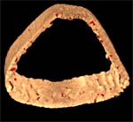
          

Cortical bone measurement
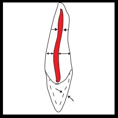
          

Tooth 3D morphological detail measurement
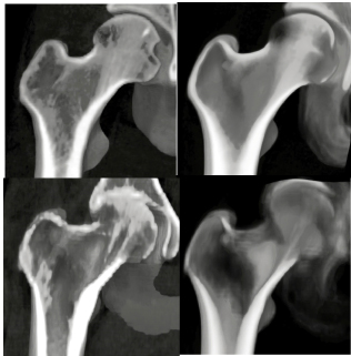
          

Medication measurement software using the VBMmethod
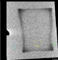
        

Measurement of enamel mineral loss
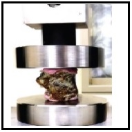
          

Finite element analysis
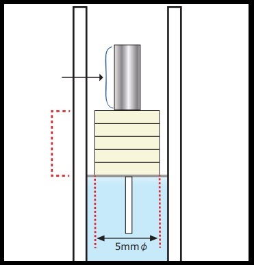
          

Bone Mineral Phantom
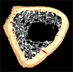
          
3D Bone morphometric measurement/option
Detailed analysis of trabecular network TRI/BON-Node StrutII
You can see the spatial distribution of plate-shaped bones and rod-shaped bones.
Thickness of trabecular bone 3D images displayed in pseudo-color show the
distribution of thickness and the state of plate-like bone resorption.
TRI series high-speed three-dimensional analysis software TRI/3D-DIF
You can automatically align
between volumes and measure bone progression between the same measurement sites.
The setting error of the measurement site can be zero.
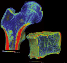
3D bone morphometrynbsp;      TRI/3D-BON-FC64
Bone quality, bone morphology, bone strength, etc. are measured using clinical X-ray
CT images and micro X-ray CT images.
It is possible to quantify the progress of osteoporosis and rheumatoid arthritis and the effect
of medication from the measurement results.
It quantifies the progress of bone resorption caused by osteoporosis and rheumatism,
and the promotion of bone formation by medication.
The handling image data size has improved dramatically.
3D bone morphometry
Automatically separates, extracts, and analyzes cancellous bone, cortical bone,
and bone marrow.

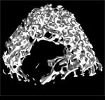
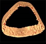
rat femur

cancellous bone

3D Node strut

cortical bone and lumen
  • Bone density BV/TV
  • trabecular width Tb.Th
  • Trabecular number Tb.N
  • Trabecular space Tb.Sp etc.
  • Trabecular surface Euler number
  • 1D Betti number
  • Trabecular surface fractal dimension
  • trabecular shape
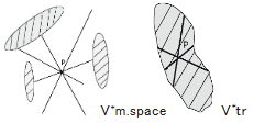
  • Trabecular structure anisotropy analysis: MIL
  • star volume
Cortical bone measurement items
  • cortical bone cross-sectional area
  • cortical bone length
  • adventitia, intima circumference
  • Moment of Inertia Area of ??Inertia, etc.
        
            star volume
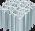
TBPf and SMI
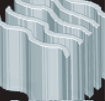
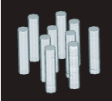
1.Concave structure          2.Plate-like                                  3.Rod-shaped
                                                                                  
TBPf < 0                                TBPf ≒ 0                                  TBPf > 0
SMI ≒ 0                                 SMI ≒ 0                                   SMI ≒ 3.0
The medullary cavity       The cancellous bone extends            It grows like a bar.
is partitioned in a     in a plate-like shape.      
honeycomb pattern   The medullary cavity is not
stretched like a bar   divided into compartments.
by platy cancellous
bones. surrounded by 
concave walls of 
small chambers     

Trabecular network structure analysis

Nd : node of multiple skeletal lines Tm : isolated endpoint Ct: Bonding point with cortical bone TV: Tissue volume to be measured
number of endpoints N.Nd N.Tm N.Ct number of skeletal lines average skeletal line length E(NdNd) E(CtNd) E(NdTm) etc.